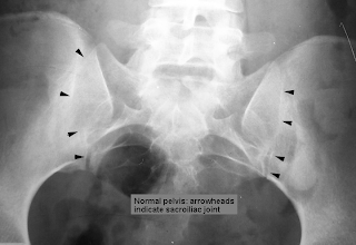
This basically means the inflammation and stiffness of a joint and in this selection I picked the spine. This is usually chronic and progressive form of seronegative arthritis. It usually affects the lumbosacral joint. Emedicine explained the Pathophysiology as; " The basic pathologic lesion of ankylosing spondylitis occurs at the entheses, which are sites of attachment to bone of ligaments, tendons, and joint capsules. Enthesopathy results from inflammation, with subsequent calcification and ossification at and around the entheses. Inflammation with cellular infiltration by lymphocytes, plasma cells, and polymorphonuclear leukocytes is associated with erosion and eburnation of the subligamentous bone. The process usually starts at the sacroiliac joints. Other enthesopathic sites include the iliac crest, ischial tuberosity, greater trochanter, patella, and calcaneum. In the paravertebral soft tissues, the lesion manifests as a formation of new bone within the outer layers of the annulus fibrosis of the intervertebral disk. The margins of the disk are invaded by hyperemic granulation tissue arising from the subchondral bone. This tissue replaces the disk fibers with new bone." I quoted the whole thing just in case I missed something and didn't explain it correctly. From what I read, there are no cures for this and it starts at young ages. They usually give pain medication for the pain. This is another one of those "well this sucks" moments. :)


Tuesday, April 21, 2009
Ankylosing Spondylitis of the spine.
Posted by Sean at 7:47 PM 0 comments
Carotid Dissection
This type of dissection can be spontaneous intracranially or extracranially. It is a significant cause of ischemic strokes which are basically mini strokes. They can be caused by major or minor trauma if there is such a thing. Most ischemic cerebral symptoms arise from thromboembolic events; therefore, early institution of antithrombotic treatment provides the best outcome. its works best if given within three hours. We have beat this horse in interventional so ill just put some cool pictures. Everyone knows that it can be caused during procedure unintentionally. THAT'S NOT A GOOD THING :)


Posted by Sean at 6:48 PM 0 comments
Tuesday, April 7, 2009
THYROID GOITER.....MY GRANNY HAD ONE ONCE
Posted by Sean at 7:43 PM 0 comments
Tuesday, March 31, 2009
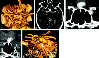
BERRY (saccular) Aneurysm
A saccular aneurysm resembles a small sack. A berry (haha yeah thats right berry) aneurysm is usually saccular. When the blood vessel weakens it can bulge out and eventually could rupture and create internal bleeding which could lead to death if untaken care of. Some options are surgery by placing a clip or stabilize the vessel with a stent if it could help. Also they can divert the blood if it isnt a very large vessel that wouldnt put the risk to high.Some rare symptoms that have happen to two patients when they described to have an unusual cause of transient ischemic attacks due to berry aneurysms of the extracranial part of the internal carotid artery. Obviously we all know what they are and i just wanted to show some cool pictures of this and just make you all aware of such a scary problem but still happens to have a cool name. ;)


Posted by Sean at 5:41 PM 0 comments
Tuesday, March 24, 2009
Nasal polyps


Posted by Sean at 5:14 PM 0 comments
Wednesday, March 4, 2009
CAVERNOUS HEMANGIOMA OF ORBIT

Cavernous hemangioma of the orbit are hamartomatous, vascular lesions, more frequent in middle aged women. They represent the most common benign primitive neoplasm of the orbit. They usually involve large groupings of blood vessels and can be removed with surgery with high risk due to locations near vital arteries. They also create blood clots that use so many platelets that the bone marrow can not keep up the production for the demand. Oral steroids can be used to reduce the size of these growths, but for serious cases surgery is the primary choice.


Posted by Sean at 7:17 PM 0 comments
Sunday, March 1, 2009
What is Acromegaly?


Posted by Sean at 6:13 PM 0 comments
Wednesday, February 25, 2009
Fibroinflammatory Pseudotumor of the Temporal Bone


Posted by Sean at 5:28 PM 0 comments
Tuesday, February 17, 2009
CRANIOPHARYNGIOMA.....yeah say that 3 times fast :p
Well for my selection of a brain pathology, I picked Craniopharygioma! What a crazy name, right? I'm going to give some suggested origins, symptoms and treatment for this disease. ENJOY!
ORIGIN: Craniopharygioma is a calcified cystic tumor located in the craniopharyngeal duct and/or Rathke cleft (pouch that usually closes in early fetal developement). Basically there are two ideas for were this come from and why it occurs. The first is the embryogenetic theory which involves CP duct and Rathke cleft. It is suggested that the remnant ectoblastic cells which in turn transform and create this tumor. The second is the Metaplastic theory which is defined as: "the residual squamous epithelium (derived from stomodeum and normally part of the adenohypophysis), which may undergo metaplasia."HAHAHAHA, yeah I'm going to quote that peach of a definition. Now I will put that in "Sean terms." Basically the cells in the general area of interest were differentiated cells and during a period of time they changed into a different type of mature cell. :)
SYMPTOMS: Some of the most common symptons include headaches, endocrine dysfunctions, and visual problems. It is said that 80% of adults complain of a not so happy or healthy sexual drive and about 90% of the men complain of impotence while women complain of amenorrhea (no menstrual period). As far as I can tell, this brain tumor is looking pretty nasty. Good news though for the young patients with this disease, most under 20 years of age survive this disease. Survival rate for those older than 65 is poor.
TREATMENT: Most stated that surgery is a very popular treatment in a attempt to remove the tumor. Also radiation therapy and chemotherapy are popular choices of treatment.
CONCLUSION: In conclusion I would like to state that even though I did make jokes and try to entertain you readers, I want to make it clear that Craniopharyngioma is a very serious brain disease and it shouldnt be taken lightly. Joking his how I deal with sometimes boring and uninteresting topics and hopefully my little ad libs were not to annoying to read. For more information on this topic I strongly suggest checking out this website: http://emedicine.medscape.com/article/1157758-overview
I got most of my information from this site. Other definitions were taken from Wikipedia. for you further enjoyment I have added some pictures! Can you tell what types of images they are?


Posted by Sean at 4:23 PM 0 comments


